Carpal hyperextension injuries are usually the result of falling from a height. Because the ligaments cannot heal to their original strength,
fusion of the joint (arthrodesis) is typically necessary to restore stable and functional limb use.
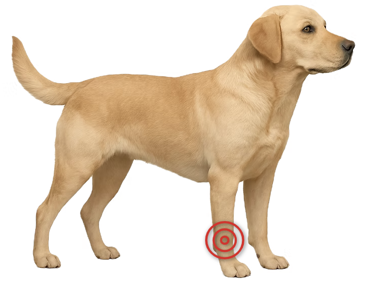
A carpal hyperextension injury occurs when the wrist joint (carpus) bends backward beyond its normal range of motion,
overstretching or tearing the ligaments that stabilize it. This usually happens when a pet falls or jumps from a height, landing with the paw flat
or even bent backward against the ground. The key supporting structures — the palmar fibrocartilage and
palmar ligaments — are essential for preventing the carpus from collapsing under weight.
Once torn, these ligaments cannot heal reliably to their original strength or length. As a result, the carpus remains unstable,
and the wrist continues to collapse during weight-bearing.
In the early stages after a carpal hyperextension injury, pets often protect the leg by holding it up or using it lightly. The wrist may appear unstable or wobbly.
As the condition progresses, the pet stands or walks with the wrist dropped closer to the ground — a position called palmigrade, meaning the paw
and wrist are flat against the floor.
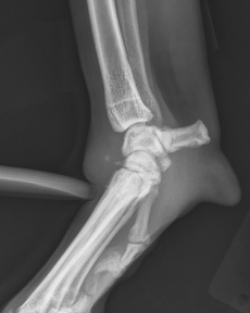
A careful orthopedic examination is critical. These injuries are sometimes missed initially, as instability can be subtle without proper manipulation.
Standard radiographs (X-rays) may not show ligament tears, so stress radiographs — which gently flex or extend the joint to reveal abnormal motion —
are necessary to confirm and localize the injury.
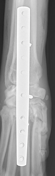
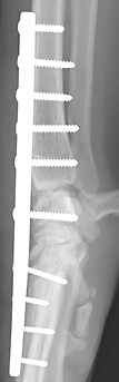
Treatment for carpal hyperextension injury is surgical, involving arthrodesis (joint fusion).
Depending on the extent of injury, this may involve:
After surgery, splinting or bandaging is required to protect the limb during healing. With proper surgical technique
and postoperative care, most dogs and cats regain strong, comfortable limb function.
Limb deformities occur when a bone grows or heals abnormally, causing it to angle, twist, turn, deviate, or shorten. These changes usually arise from growth plate problems due to genetics (as in Dachshunds and other chondrodystrophic breeds), trauma that damages a growth plate, or bones that heal improperly after a fracture (malunions).
Depending on severity, deformities may cause visible limb dysfunction and, if the joints are affected, can lead to arthritis. In some dogs, however, mild deformities cause little to no noticeable problem.
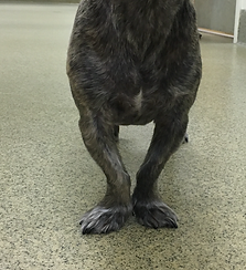
Bilateral antebrachial (forelimb) deformities in a dog.
Limb deformities are usually obvious when there is visible bending or twisting of a leg, which may gradually worsen over time. Different bones can be affected, and deformities may lead to limping — either from abnormal mechanics or from soreness in the affected limb.
Evaluation begins with a comprehensive orthopedic examination. Your pet’s gait is observed to assess limb function and balance. The affected limb is then carefully examined to check alignment, measure length, and detect any pain or joint changes.
Radiographs (X-rays) are the most important initial diagnostic tool. They are taken from multiple angles and often include the opposite limb for comparison. In more complex cases, a CT scan may be performed to create a 3D model of the bones, allowing precise surgical planning. If surgery is anticipated, routine bloodwork and urinalysis are also done to ensure your pet is healthy for anesthesia.
Not all limb deformities require surgery. Mild deformities that cause little or no discomfort can often be monitored over time, especially if your pet is functioning well.
In young, growing dogs, carefully timed procedures such as partial bone excision (ostectomy) may allow the limb to correct itself naturally as growth continues. For more significant deformities — especially those causing pain, limping, or risk of joint degeneration — a corrective osteotomy is usually recommended. In this procedure, the bone is precisely cut, realigned to restore normal mechanics, and stabilized using plates, screws, or external fixators.
In advanced or highly complex cases, 3D-printed surgical guides and detailed pre-operative computer modeling may be used to plan the correction with extreme accuracy.
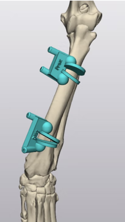
Computer model showing custom guides to assist with limb deformity correction.
The outlook for recovery from a limb deformity varies widely depending on severity, age, and overall health. Mild cases can have excellent long-term outcomes without intervention, while severe deformities may require complex surgical correction.
Successful outcomes rely on accurate surgical technique, proper implants, and careful post-operative management. With expert planning and follow-up, most dogs and cats regain comfortable function and improved limb alignment after corrective surgery.