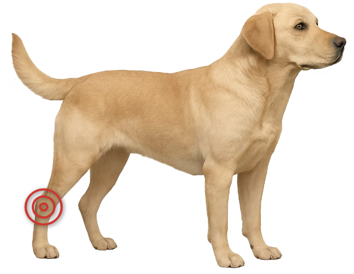
Hock (ankle) problems are an important cause of lameness in dogs and cats, as it is prone to improper development as well as traumatic injury.
Osteochondritis dissecans (OCD) — a progression of osteochondrosis — is a joint condition in young, growing dogs where cartilage fails to form properly, leading to pain and lameness. It can occur in several joints, most often the shoulder, elbow, stifle, or hock.
Osteochondrosis is a developmental disorder in which the normal process of cartilage turning into bone (endochondral ossification) is disrupted in a growing dog. When a section of this abnormal cartilage cracks or separates from the underlying bone, it forms a loose or partially attached flap — a stage called osteochondritis dissecans (OCD). This flap can cause joint irritation, inflammation, and mechanical interference with movement, leading to pain and lameness.
OCD is more common in large and giant breeds, and genetics play a major role. Other contributing factors may include rapid growth, diets high in calcium or energy, repetitive joint stress, and hormonal influences.
Osteochondrosis itself often causes no symptoms because the abnormal cartilage remains intact and does not irritate the joint. When it progresses to OCD, the detached or partially attached flap causes inflammation and irritation, leading to persistent pain and limping.
Signs typically appear in young, growing dogs between 6–18 months of age. It can also be a cause of lameness in older dogs, especially once secondary arthritis develops.
OCD lesions are usually visible on high-quality radiographs (X-rays), particularly in the shoulder, elbow, stifle, or hock. CT scans are sometimes recommended to better define the size, shape, and exact location of the defect, which assists with surgical planning. Arthroscopy allows direct visualization and confirmation of cartilage flap formation.
Osteochondrosis without pain does not require surgery and can be monitored over time. However, OCD almost always causes persistent lameness, and surgical removal of the cartilage flap generally leads to significant improvement, though outcomes vary depending on the joint.
Surgery can be performed using either an open approach or minimally invasive arthroscopy. Arthroscopy typically allows for faster recovery, smaller incisions, and less postoperative discomfort. In cases where the lesion is large and leaves a substantial defect, resurfacing techniques such as grafting or synthetic implants may be used to restore a functional joint surface.
Hock (ankle) trauma in dogs and cats can involve ligaments, tendons, and multiple bones.
The joint is complex, with several stacked levels, any of which can be injured.
The hock (ankle) is a complicated joint made up of several smaller joints stacked together.
The top joint between the shin bone (tibia) and the main ankle bone (talus) allows most of the movement,
while the lower joints move much less but still play an important role in stability.
Strong collateral ligaments help keep the hock steady, but the area has very little muscle or soft tissue padding,
which means the bones, tendons, and skin are more exposed to injury.
Because of its structure and location, the hock can be injured in many different ways. Twisting the joint, getting a leg caught,
or being hit by a car can damage ligaments, tendons, or bones, and the hock is somewhat prone to fractures
and dislocations. Its position near the ground and limited soft tissue coverage also make it vulnerable to
wounds, especially shear injuries where the skin is scraped or torn.
With major hock injuries, dogs or cats usually show obvious lameness and may refuse to use the leg at all.
Swelling is often visible around the joint, and the animal may pull the leg away or react when the area is touched.
Localizing an injury to the hock is usually straightforward, but identifying exactly which structures are damaged requires
a detailed examination. The veterinarian will carefully feel and move the joint, often “stressing” it in specific
directions to check for abnormal looseness or pain, which can point to injuries of particular ligaments or supporting tissues.
Radiographs (X-rays) are taken in both neutral and stressed positions to assess bone alignment, detect fractures,
and reveal instability. In more complex or unclear cases, a CT scan may be recommended for a highly detailed view
of the bones and joint surfaces.
Applying controlled “stress” to the joint can help demonstrate ligament rupture on radiographs.
Major injuries to the hock’s key structures often require surgery. Depending on the type and severity of damage,
treatment may involve:
Postoperative immobilization with a splint or external frame is frequently needed to protect the repair while it heals.
Successful outcomes depend on prompt intervention and a surgeon with detailed knowledge of hock anatomy, joint mechanics,
and the unique challenges of this complex, multi-level joint.
Collateral ligament reconstruction is a common surgical approach when these key stabilizing structures are damaged.
Prognosis is widely variable and depends on the specific structures injured, the severity of damage, the accuracy of surgical repair,
and how quickly treatment is performed. Injuries limited to a single ligament or bone generally have a better outlook than those
involving multiple levels of the joint or extensive soft tissue loss.
Fractures that extend into the joint surface, severe dislocations, or cases requiring arthrodesis tend to carry a more guarded prognosis.
Long-term outcomes can also be affected by the development of secondary arthritis, which is common after significant hock trauma.
Info Coming Soon
Osteoarthritis is an extremely common problem in both dogs and cats. In some animals, it is well tolerated and causes minimal issues, while in others it is debilitating. Prevention and treatment recommendations are highly individualized.
Osteoarthritis, also known as degenerative joint disease (DJD), arthritis, or osteoarthrosis, involves thinning and wearing of cartilage. It also affects surrounding joint tissues, including bone, ligaments, the joint capsule, and nearby muscles. As degeneration progresses, the underlying bone becomes exposed and both soft and hard tissues can thicken. The joint then loses its ability to glide smoothly through its normal range of motion.
In dogs, osteoarthritis is typically secondary — developing as a consequence of another joint problem such as ligament injury, fracture, or developmental disorders like elbow or hip dysplasia. Because secondary OA is so common, identifying and addressing the underlying problem is critical to proper treatment planning.
Classic symptoms of osteoarthritis include:
In cats, signs are often more subtle — such as less movement around the home, avoiding touch, or reduced grooming habits. Importantly, many animals with osteoarthritis show no symptoms at all.
A careful orthopedic examination is crucial. Affected joints may show thickening, reduced range of motion, pain, or crepitus (a crunching sensation). Multiple joints may be involved, so a full evaluation is essential.
It’s also important to determine underlying causes. For example, instability in an arthritic stifle can indicate secondary arthritis due to a cranial cruciate ligament rupture.
Radiographs (X-rays) are essential for confirming osteoarthritis, identifying its cause, and gauging severity. Advanced imaging such as CT or MRI can reveal more detail about bone and soft tissue changes. Arthroscopy allows direct visualization of the joint interior and can be used both to confirm diagnosis and treat certain causes of secondary OA.
The goal of treatment is to reduce pain, improve movement, and slow further joint damage. Osteoarthritis is often managed medically, including anti-inflammatory medications, weight control, joint supplements, and physical therapy. Many pets stay comfortable for years with these measures alone.
Surgical options may be recommended for different reasons:
The best approach depends on which joint is affected, disease severity, your pet’s overall health, and lifestyle needs.
Most dogs can enjoy many years of good quality life with the right management plan. The condition’s course varies — some dogs remain asymptomatic, while others develop significant mobility issues that may progress with age.
Early diagnosis and proactive care — including weight management, tailored exercise, pain control, and, when appropriate, surgical intervention — can slow progression and dramatically improve comfort and mobility.
At Animal Orthopaedic Clinic of Florida, every patient benefits from unmatched expertise. Our head clinician is an internationally recognized leader and researcher in veterinary orthopaedic surgery. Using evidence-based, individualized care, we are dedicated to giving your dog the best chance at long-term, pain-free mobility.
Longo F, Castelli E, Lewis DD, Hudson CC, Kim SE, Pozzi A. Minimally invasive tarsal arthrodesis in 15 dogs. Vet Surg 2025; 54:129–140.
Evers JS, Kim SE*. Use of a bone-to-tendon plate to stabilize a comminuted calcaneus fracture in a dog. Vet Surg 2022; 51:859–863.
Franklin SP, Stoker A, Murphy S, Kowaleski MP, Gillick M, Kim SE, Karlin M, Cross AR, Cook JL. Outcomes associated with osteochondral allograft transplantation in dogs. Front Vet Sci 2021; 8:759610.
Lazarus MA, Kim SE*, Lewis DD, Johnson MD. Intra-articular injection of a dextran polymer combined with antibiotics for bacterial infective arthritis in companion animals: 15 cases. Vet Comp Orthop Traumatol Open 2021; 4:e92.
Pozzi A, Lewis DD, Hudson CC, Kim SE, Castelli E. Percutaneous plate arthrodesis. Vet Clin North Am Small Anim Pract 2019; S0195-5616(19)30128-7.
Hudson CC, Kim SE, Pozzi A. Percutaneous pinning for fracture repair in dogs and cats. Vet Clin North Am Small Anim Pract 2019; S0195-5616(19)30132-9.
Boekhout CL, Kim SE, Pozzi A, Cross AR. Closed reduction and fluoroscopic-assisted percutaneous pinning of 42 physeal fractures in 37 dogs and 4 cats. Vet Surg 2017; 46:103–110.
Dunlap AE, Kim SE*, McNicholas WT Jr. Biomechanical evaluation of a non-locking pre-manufactured loop suture technique compared to a three-loop pulley suture in a canine calcaneus tendon avulsion model. Vet Comp Orthop Traumatol 2016; 29:131–135.
Kim SE*, Hudson CC, Pozzi A. Percutaneous pinning for fracture repair in dogs and cats. Vet Clin North Am Small Anim Pract 2013; 42(5):963–974.
Pozzi A, Lewis DD, Hudson CC, Kim SE. Percutaneous plate arthrodesis in small animals. Vet Clin North Am Small Anim Pract 2012; 42(5):1079–1096.