
ANIMAL

ORTHOPAEDIC

CLINIC

OF FLORIDA

Muscle and Tendon Injury

Home / Our Services / Muscle and Tendon Injury

Muscle and tendon injuries in dogs and cats can seriously affect limb function. While many result from trauma, others may occur spontaneously due to underlying joint or tissue conditions.

What are they?

Muscles and tendons work together to make movement possible. Muscles generate force, and tendons act like strong ropes attaching muscles to bones. Injuries can occur in several ways:

  • Bruises or low-grade strains – Overuse or sudden overstretching can cause soreness, swelling, or small fiber tears.
  • Partial or complete tears – If fibers rupture, the muscle or tendon may stop functioning properly, causing limping or inability to bear weight.
  • Cuts (lacerations) – Sharp injuries can slice through tendons or muscles, especially on the lower legs.
  • Chronic problems – Repeated stress can weaken tissues over time, leading to tendinopathy.

Some injuries are mild and heal with rest, while others are more serious and require surgery to restore function. If the injured structure is critical for weight-bearing — such as the Achilles tendon — the problem is often much more noticeable.


Symptoms

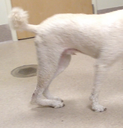
Signs depend on the severity and location of the injury. Mild injuries may cause only short-term symptoms, while more severe damage results in persistent limping. Swelling or tenderness may occur, but are not always obvious. After certain tendon injuries (e.g., the Achilles complex), the limb may appear to collapse during weight-bearing.

A dog with a ruptured Achilles tendon — note the ‘dropped’ hock.


Tests

Diagnosis begins with a thorough hands-on exam. Your veterinarian will observe how your pet walks, feel the affected area for pain and swelling, and move the joint through its range of motion. These initial steps often provide key clues.

Additional imaging may be recommended to confirm the diagnosis or rule out other issues:

  • X-rays – Identify bone fractures or bone spurs where tendons attach. While tendons themselves aren’t well visualized, surrounding changes can provide clues.
  • Ultrasound – Excellent for visualizing tendons and muscles, detecting fiber tears, fluid buildup, or tissue thickening from repeated strain.
  • MRI – Used for complex cases, especially in areas like the shoulder where multiple structures overlap.
  • Joint tap or contrast X-ray – Sometimes used to assess tendon involvement in shoulder conditions.

Mild strains may not show clear changes on imaging. In these cases, improvement with rest and controlled rehabilitation helps support the diagnosis.


Treatments

Treatment is tailored to the injured structure and severity:

  • Conservative care: Rest, controlled activity, anti-inflammatory medication, and structured rehabilitation.
  • Immobilization: Splints or bandages may be needed for several weeks to protect healing tissues.
  • Targeted therapies: Options like shockwave therapy or selective corticosteroid injections can be beneficial in some tendinopathies.
  • Surgery: Required for significant tears or lacerations. Precise tendon repair techniques are used to minimize gaps, and supportive bandaging is typically required afterward.

Prognosis

Outcomes depend on which muscle–tendon unit is affected, the severity of the injury, how quickly treatment begins, and the quality of rehabilitation. Many pets return to comfortable function with proper care. Injuries to major weight-bearing tendons require longer recovery times. Chronic contracture disorders and certain myopathies carry a more guarded prognosis — early detection and proactive management are key.

Medical content reviewed by: S. Kim

Why AOCF?

At Animal Orthopaedic Clinic of Florida, every patient benefits from unmatched expertise. Our head clinician is an internationally recognized leader and researcher in veterinary orthopaedic surgery. Using evidence-based, individualized care, we are dedicated to giving your dog the best chance at long-term, pain-free mobility.

Muscle and Tendon Research by AOCF Surgeons +

Maki LC, Kim SE*, Winter MD, Kow K, Conway JA, Lewis DD. Compartment syndrome associated with expansile antebrachial tumors in two dogs. J Am Vet Assoc 2014; 244:346-351.

Dunlap AE, Kim SE*, McNicholas WT Jr. Biomechanical evaluation of a non-locking pre-manufactured loop suture technique compared to a three-loop pulley suture in a canine calcaneus tendon avulsion model. Vet Comp Orthop Traumatol 2016; 29:131-135.

Evers JS, Kim SE*. Use of a bone-to-tendon plate to stabilize a comminuted calcaneus fracture in a dog. Vet Surg 2022; 51:859-863.

de Moya K, Kim SE*. Radiographic evaluation of patellar tendon length following corrective surgical procedures for medial patellar luxation in dogs. PLoS ONE 2020; 15:e0238598.